AI-supported dermatology

We accelerate the diagnosis process from dermatological findings with our artificial intelligence supported systems.

We accelerate the diagnosis process from dermatological findings with our artificial intelligence supported systems.

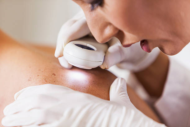
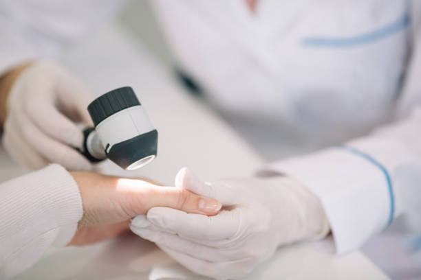
‍Cancer can cause death if not diagnosed and treated in a timely fashion. Skin cancer is a common type of cancer, as more than three million Americans are diagnosed with skin cancer each year. Melanoma is the most dangerous type of cancer and tends to spread rapidly. Unaided visual inspection of expert dermatologists yields diagnostic accuracy of about 60%. Dermoscopy is a noninvasive imaging technique for the microscopic examination of pigmented skin lesions, placing a high-resolution magnifying imaging device in contact with the skin. Lighting is controlled, and a liquid interface or polarization filter is applied to remove surface skin reflectance, exposing underlying layers of skin for inspection. Dermoscopic imaging has been shown to improve recognition accuracy and provides accuracy between 75%-84%. Dermoscopy results in lower diagnostic accuracy if the physician does not recognize or correctly interpret the significance of structures.

  • Industry
    Healthcare
  • Duration
    12 months
  • Team size
    12
  • Technologies
    Healthcare

Our Technology

Our Artificial Intelligence (AI) supported dermatology solution uses a machine-learning algorithm based on ensembling deep learning models. Deep models have been trained using a dermoscopic imaging database containing tens of thousands of examples that have confirmed diagnosis and assessment by dermatologists.

Different from the traditional ABCDE (asymmetry, boundary, color, diameter, and change over time) features, our AI-based solution distinguishes between benign and malignant tumors by considering thousands of deeply extracted features.

Eight categories of skin cancers Melanoma (MEL), Actinic Keratosis (AK), Benign Keratosis (BK), Basal Cell Carcinoma (BCC), Dermatofibroma (DF), Squamous Cell Carcinoma (SCC), Melanocytic Nevi (NV), and vascular Lesions (VASC) are automatically detected.

How to use our technology?

  • Take a dermoscopic photo simply focusing on a lesion using a mobile phone with a lens attached on.
  • Sent your photo to the server via our app.
  • Learn your risk score assessed by the AI-based algorithm.

Benefits of AI-based dermoscopic assistance system

  • Our solution turns the subjective assessment made by the specialist into an objective.
  • Provides high classification and repetition accuracy in less than ten seconds.
  • No need for high-cost hardware for image capture.
  • Accessible any time and anywhere.

“Amatis is changing our business with all sorts of new technics. With their end-to-end solutions, we can offer our customers a better product and reduce our production time. We are back in the top-notch medical systems with the AI and ML algorithms incorporated in our analysis software.”

Robert Den Engelsman
CEO
Fysiologic

Your digital partner in healthcare

Get in Touch

Related cases

Healthcare - AI & Big Data Analytics

ECG Analysis with AI

Healthcare

Telehealth

Healthcare

Sleep apnea

Healthcare

Flow cytometry